Vascular ultrasound platform

Report, learn and practice vascular duplex in one place.

iDoppler brings together what a vascular lab needs: structured reports in your own template, vascular mapping, a technical review of your images, and a complete course with a question bank. Built by a vascular surgeon, for people who scan every day.

Phone, tablet and computerEnglish · Español · PortuguêsNothing to install
Dr. Vinicius Majdalani, in a navy blazer and black-framed glasses
Vinicius Majdalani, MD Senior RVT · RPVI · ABVM 18 years in vascular ultrasound Board-certified vascular surgeon (Brazil)

Why iDoppler exists

Anyone who scans knows three bottlenecks of the daily routine.

The report

Typing eats into scanning time.

Diameters, reflux, segments and sides. Every venous study turns into minutes of typing after the patient has left the room.

The image

Technique rarely gets a second opinion.

Angle, scale, gain and color box determine the measurement. Almost nobody has someone to review the image after acquisition.

Training

The content is scattered.

Physics, criteria and technique are spread across textbooks, consensus papers and loose lectures, with no clear path from physical principle to final report.

Reports

From dictation to PDF, in your own template.

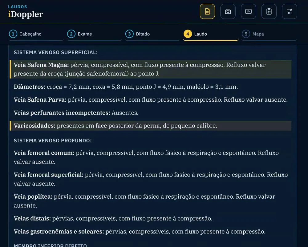
You start from your normal report and enter only the findings. iDoppler organizes the text, highlights every changed line for your review, and draws the vascular map from the final report.

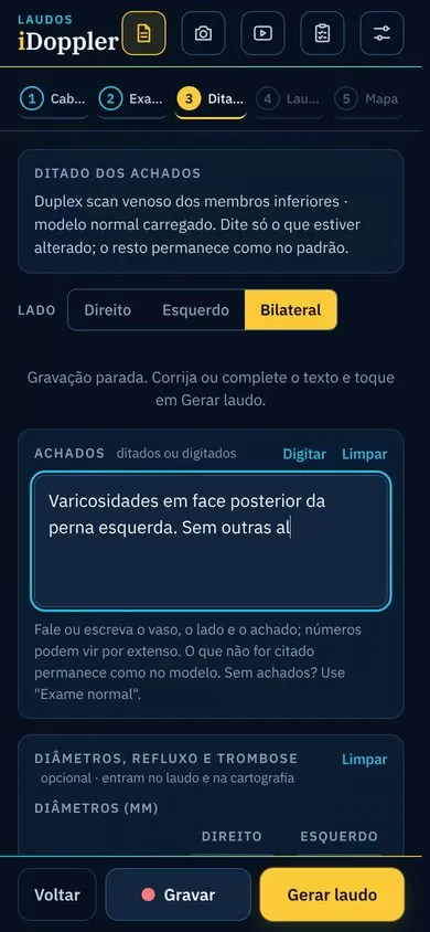
  1. Dictate or type the findingsChoose the exam and the side. Tap Record and say only what is abnormal.
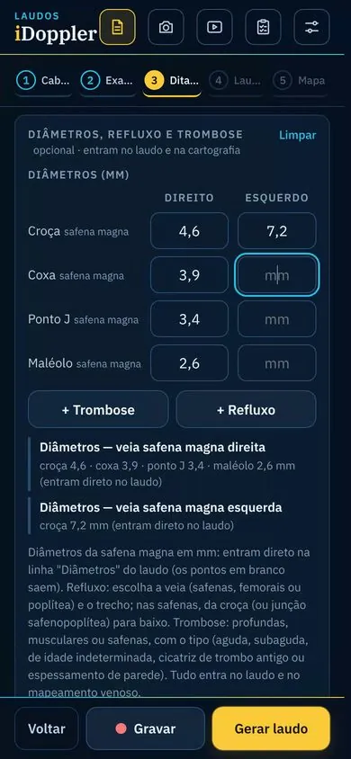
  2. Use buttons when it is fasterSaphenous diameters in a table; reflux and thrombosis with side, vein and segment in one tap.
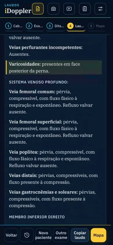
  3. Review the generated reportThe text follows your template, with the changed lines highlighted. The final word is always yours.
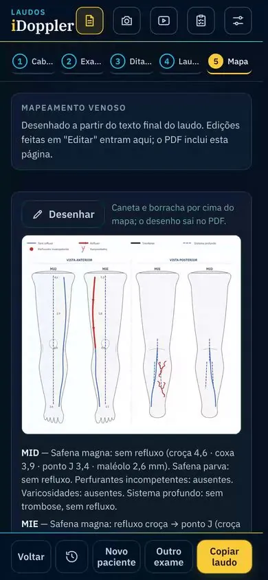
  4. Deliver it with a vascular mapA map drawn from the report, editable with a stylus, and a PDF with your logo and signature.
iDoppler screen: dictating the findings of a lower-extremity venous duplex iDoppler screen: saphenous diameter table and reflux and thrombosis buttons iDoppler screen: generated report with the changed lines highlighted iDoppler screen: vascular map drawn from the report
Vascular map on the phone

Real app screens with sample data, shown in the Portuguese interface. The app also runs in English and Spanish.

iDoppler · Reports
The same report open on a computer, with the changed lines highlighted
Start on the phone, finish on the computerThe report is saved to your account and shows up on any device, along with your previous reports.
Paste it into your systemCopy it into your clinic's records, or generate a blank PDF with margins for your letterhead.
Your template, your wordsThe normal report templates for each exam can be edited in Settings.
Signature and brandingLogo, footer, address and scanned signature go into the PDF.
Arterial and venous examsCarotid and vertebral, upper- and lower-extremity arterial and venous, aorta and iliacs, renal and more.
Three languagesInterface in English, Spanish and Portuguese.
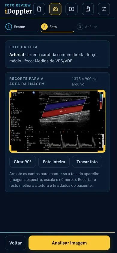
Foto Review: photo of the scanner screen, cropped to the image area
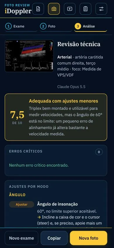
Foto Review: technical review with a 7.5 score and adjustments by mode

Foto Review

A second opinion on the technique behind your image.

Take a photo of the scanner screen and tell iDoppler the vessel and the focus of the review. In seconds you get a score and what to adjust in each mode, B-mode, color and spectral, so your next acquisition is better.

7.5out of 10

Adequate, with minor adjustments. Right common carotid artery, mid segment. Focus: PSV/EDV measurement.

Angle of insonation · adjustColor box · adjustSpectral scale · okBaseline · ok

Foto Review assesses the technical quality of the acquisition. It does not interpret the exam or make a diagnosis. On a computer, the photo arrives from your phone through a QR code.

DOPPLER Course · Early 2027

From physical principle to final report.

The course Dr. Majdalani teaches to specialist physicians, now inside iDoppler. It starts with ultrasound physics, moves through scanner settings and reaches the criteria for each vascular territory, based on the main Brazilian, American and European consensus documents. All lessons have English and Spanish subtitles. Fully narrated English versions, with translated slides, are being released module by module, starting with ultrasound physics.

160+video lessons
3languages: subtitles in English and Spanish
500+original questions
  • Pick up where you left off, on any device, with progress for each module.
  • Clickable chapters and notes pinned to the minute of the video.
  • PDF summaries of the lessons, to review before an exam or a shift.
  • Question bank and Foto Review included in the Course plan.

Physics and instrumentation

34 lessons

Physical principles, the Doppler effect, B-mode and color settings, spectral analysis, direct and indirect signs, and spectral settings.

Carotid and vertebral

Cerebrovascular

Technique, stenosis criteria, vertebral arteries, other carotid diseases, endarterectomy and follow-up, ophthalmic artery and cervical veins.

Peripheral arterial

Lower and upper extremity

Peripheral artery disease, other arterial diseases, post-intervention follow-up and the upper-extremity arterial exam.

Venous

Lower and upper extremity

Chronic venous insufficiency, deep vein thrombosis, post-procedure follow-up, hemodialysis arteriovenous fistula and the upper-extremity venous exam.

Abdomen: aorta and renal arteries

Growing

New aorta and renal artery lessons are added as they are recorded.

Extra: ultrasound-guided venous access

Extra

Ultrasound fundamentals for venous access, internal jugular and subclavian anatomy, sterile preparation and real-time puncture, in-plane and out-of-plane, with 3D animations.

Extra: anatomy of the transducer

6 lessons

How the transducer works, and how to choose and position it for each exam.

Questions

Train like it's exam day. Understand every option.

More than 500 original questions, from intermediate to advanced, in English, Spanish and Portuguese. Build blocks by subject and territory, in tutor mode or as a timed exam.

Every option explainedWhy the right answer is right and why each wrong one is wrong, with the reference.
How your peers answeredThe percentage of physicians who chose each option.
Your performanceScore by subject and territory, percentile among physicians and time per question.
Pause and resumeStop a block halfway and pick it up later, right where you left off.
Physics · Spectral DopplerTutor mode

In the same medium, the operator increases the transducer frequency. What happens to the propagation speed of sound?

AIt increases in the same proportion22%
BIt decreases in the same proportion9%
CIt does not change: it depends only on the medium61%
DIt depends on the focal depth8%

Propagation speed depends on the stiffness and density of the medium. If frequency goes up, wavelength goes down, and c = f × λ stays the same.

Sample question written for this page. Percentages are illustrative.

Plans

Choose what fits where you are.

To report faster, choose Practice. To study and prepare for exams, choose Course. To get to know the platform, start for free.

Get started

Free

Your normal report templates always at hand, on your phone and your computer.

  • Normal report templates for the main vascular exams
  • Manual report editing
  • Vascular map to draw on with your finger, a stylus or a mouse
  • English, Spanish and Portuguese
  • No dictation or generated report
Create free account
Coming soon · For reporting

Practice

For physicians who scan every day and want to report in less time.

  • 150 reports per month, by dictation or typing
  • Changed lines highlighted for review
  • Vascular map generated from the report
  • PDF with logo, footer and signature
  • Foto Review: 10 reviews per month, up to 5 questions per photo
  • Reports saved and synced across devices
Notify me at launch
Early 2027 · For studying

Course

For those training in vascular ultrasound or preparing for board and credentialing exams.

  • DOPPLER Course: 160+ video lessons with English subtitles
  • Chapters, notes and PDF summaries
  • Questions: full bank, unlimited
  • Tutor mode, timed exams and statistics
  • Foto Review: 10 reviews per month, up to 5 questions per photo
  • Reports not included
Notify me at launch

Want both? The PRO plan will combine Practice and Course, with higher limits: 300 reports and 15 Foto Reviews per month.

Notify me at launch

Practice coming soon. Course in early 2027. Free account holders hear first.

Coming soon

Something new that will be a game changer in vascular ultrasound education.

We are preparing something that changes how vascular duplex is learned and practiced. iDoppler account holders will be the first to know.

Save my spot on the list
Portrait of Dr. Vinicius Majdalani

Who built it

Vinicius Majdalani, MD

Vascular surgeon and vascular ultrasound specialist with about 18 years of duplex practice, faculty in a vascular surgery residency program and director of the DOPPLER course for specialist physicians in Brazil, where he is board-certified in vascular surgery and in vascular ultrasound.

In the United States he holds the Registered Vascular Technologist (RVT) and Registered Physician in Vascular Interpretation (RPVI) credentials, and is certified by the American Board of Vascular Medicine (ABVM).

Senior RVTRPVIABVM
I built iDoppler to solve what I lived in the scanning room: less time typing, more time with the patient, and a clear path for those who are learning.

Frequently asked questions

Before you start

Does iDoppler make a diagnosis?

No. iDoppler is a tool for transcribing, formatting and mapping reports, and for education. The physician performs the exam, makes the decisions and reviews every line of the report. Foto Review assesses only the technical quality of the image.

Do I need to install anything?

No. iDoppler opens in the browser of your phone, tablet or computer. Your reports are saved to your account and show up on all your devices.

Can I use it with my clinic's reporting system?

Yes. Copy the finished report and paste it into the system you already use, or generate a blank PDF with your letterhead margins. If your system prints the report, print only the vascular map.

What about patient data?

The patient's name is optional, so you can create reports without identifiers. Reports are stored in your account, accessible only to you, as described in the platform's terms of use and privacy policy.

Is the app in English?

Yes. The interface, the report templates and the question bank are available in English, Spanish and Portuguese. All course lessons have English and Spanish subtitles, and narrated English versions with translated slides are being released module by module.

Can I start for free?

Yes. The free account gives you the normal report templates and the vascular map to draw on. When you want dictation, generated reports, the course or the questions, just switch plans.

Start today

Your next vascular report can start here.

Create your free account in under two minutes and be the first to know when Practice and Course open.

Create free account Instagram @idoppler.app
Email:contato@idoppler.com.br